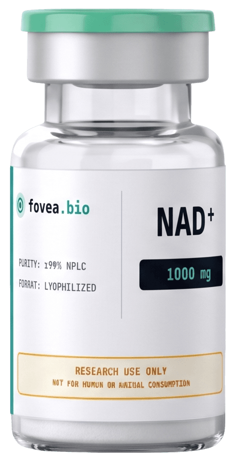
NAD⁺
β-Nicotinamide Adenine Dinucleotide (NAD⁺)
- ≥99% HPLC
- LC-MS Verified
- Tested
- Vacuum-Sealed
- Cold-Chain Ready
The central energy-transfer molecule every cell runs on. Retinal cells burn through enormous amounts, and it is the fuel for the sirtuin repair enzymes. In laboratory assays, it is used to study cellular repair pathways, optimize mitochondrial ATP production, and combat age-related metabolic decline.
Schematic only : not to scale
Relevance to Vision Research
Foveal photoreceptors and retinal ganglion cells are among the most energy-hungry cells in the body, making their NAD⁺ supply a central variable in retinal bioenergetics research.
Where It Acts : Pathway Map
11 structures targetedAn animated map of the visual pathway, from the eye through the brain to the systemic processes of aging. Highlighted nodes mark the structures NAD⁺ is studied against.
Illustrative research map : highlighted structures reflect the published in-vitro / preclinical literature, not human outcomes.
Key Performance Benefits
Scientifically supported advantages for peak performance and recovery.
Maximizes ATP Energy Production
Serves as the critical fuel for mitochondria to convert nutrients into massive cellular energy, unlocking explosive power and peak physical performance.
Extends Athletic Endurance
Rapidly replenishes redox cofactors depleted during high-intensity training, significantly delaying muscle fatigue and extending stamina.
Optimizes Metabolic Repair
Powers essential repair enzymes (sirtuins and PARPs) to protect DNA, optimize metabolism, and accelerate muscular recovery between sessions.
Disclaimer: NAD+ should be used responsibly. These statements have not been evaluated by the FDA. Not intended to diagnose, treat, cure, or prevent any disease.
What the Research Shows
2 references ↓Protected retinal ganglion cells[1]
NAD⁺ repletion prevented RGC loss in a landmark aged-mouse glaucoma study.
The cellular fuel cofactor[2]
Essential carrier in the energy chain that retinal cells depend on most.
Powers the repair enzymes[2]
The substrate sirtuins and PARPs consume; tissue levels decline with age.
Sharper energy metabolism[2]
Central to converting nutrients into the ATP that retinal cells consume rapidly.
Summaries of observations reported in published in-vitro and preclinical research. These are research-model findings, not established human outcomes, and not claims of benefit, safety, or efficacy. Supplied for laboratory research use only.
Literature Protocol Reference
Preclinical and clinical trial parameters documented in published literature. Reconstituted reference materials are designed for laboratory analysis and in-vitro assays.
Research Literature Notes
Subcutaneous (SC) protocols utilize 25 mg to 100 mg daily (50 mg/day is standard; 1 vial of 1000 mg provides a 20-day course at 50 mg/day). Slow IV infusion protocols utilize 250 mg to 500 mg diluted in 250 mL of NaCl 0.9% infused over a minimum of 2-4 hours. Reconstituted NAD+ is highly unstable and must be stored refrigerated and used within 24-48 hours. Never mix with other peptides.
Why It Is Used
NAD⁺ is a small, highly polar dinucleotide that is fully water-soluble and exceptionally well-characterized, making it a clean analytical reference standard. It is light- and moisture-sensitive in solution, so the lyophilized, amber-sealed format is the appropriate handling state. The in-house conformer ensemble is used mainly to calibrate docking grids against NAD⁺-binding oxidoreductase pockets.
Objective structural / physicochemical opinion. Not medical advice.
Related Scientific Reports
Read the full literature review on NAD⁺Retinal Ganglion Cell Bioenergetics: NAD⁺ Flux and the Foveal Mitochondrial Load
A structured synthesis of the published literature on energy demand in the foveal cone and retinal ganglion cell (RGC) axis, the role of the NAD⁺/NADH redox pair, and reported RGC protection following NAD⁺ repletion in optic-neuropathy and glaucoma models. Presented as a neutral reference survey; no human outcome or product effect is asserted.
Photoreceptor Phototoxicity and Antioxidant Peptide Strategies: A Structural Survey
A structural survey of high-energy-visible (HEV) photo-oxidative stress pathways in photoreceptor and RGC mitochondria, mapped against the reported antioxidant and membrane-stabilizing properties of the catalogued short peptides. Combines a literature map with in-house physicochemical descriptors. Reference survey only.
Documented References
- [1]
Williams PA, et al. (2017) Vitamin B3 modulates mitochondrial vulnerability and prevents glaucoma in aged mice.
Science
View on PubMed - [2]
References point to published, third-party scientific literature, provided for research context. Citation of a study is not an endorsement of any use of this material.
Molecular Identity
- CAS Number
- 53-84-9
- Molecular Formula
- C₂₁H₂₇N₇O₁₄P₂
- Molecular Weight
- 663.43 g/mol
- Purity
- ≥ 99.0% (HPLC, area)
- Format
- Lyophilized powder, vacuum-sealed amber vial
Analytical Specification
Research Context
NAD⁺ is the central pyridine-nucleotide redox cofactor mediating electron transfer in glycolysis, the TCA cycle, and oxidative phosphorylation, and is a consumed substrate of the sirtuin (SIRT1–7) and PARP enzyme families. The published literature documents a decline in tissue NAD⁺ pools with age and metabolic stress. Catalogued here as an analytical reference standard for cofactor-dependent enzyme assays.
Reference conformer ensemble generated locally (RDKit ETKDGv3 + MMFF94s) for docking-grid calibration against NAD⁺-binding oxidoreductase pockets.
Laboratory Handling
Soluble in laboratory-grade water and standard aqueous buffers. Solubility and stability data provided for in-vitro assay design only.